Structure and Function of Fibroblast Cells
Interspersed between the collagen fibers of dense connective tissue and the interstitial spaces in epithelial tissues are fibroblast cells. These cells are responsible for collagen production and serve to maintain the extracellular matrix and stroma in connective tissues. Fibroblasts do this by secreting compounds that serve as precursors to components of the extracellular matrix, which go on to form collagens, glycosaminoglycans, glycoproteins and reticular and elastic fibers. The extracellular matrix is important because its composition determines the physical properties of the connective tissue.
The fibroblast population exists as cells in multiple stages of differentiation. Fibroblast cells look long and flat with several elongated processes protruding from the body of each cell. The processes are the result of a branched cytoplasm, which surrounds an elliptical, spotted nucleus. The nucleus can have one or two oval nucleoli which appear as dark spots under the microscope. Cytoplasm components include mitochondria near the nucleus and a small lipid droplet.
Fibroblasts aid in maintaining the structure of connective tissues by the secretion of precursors of the extracellular matrix, which is primarily a mixture of fibers and the ground substance. Fibroblasts, along with related connective tissues help sculpt the majority of an organism and can migrate slowly over a substrate as individual cells. Although they often align in parallel when crowded, fibroblasts show dissimilar phenotypes in different anatomical locations. Unlike epithelial cells lining body structures, fibroblasts are not restricted to a basal lamina on one side and do not form flat mono-layers.
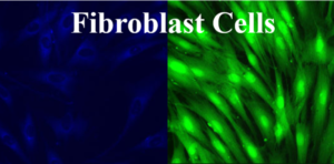
Copyright picture from altogen.com. Reproduced with permission from Altogen Biosystems.
Tissue matrix is made almost entirely of collagen, which is the most abundant protein in mammals. Between collagen fibers of connective tissue are fibroblasts, which produce a collagen subunit called tropocollagen. Tropocollagen is used to make larger collagenous aggregates.
Fibroblasts produce polysaccharides and glycoproteins, a gel-like material that surrounds collagen fibers of dense connective tissue. This “extracellular matrix” contributes to the structure of ligaments and tendons. In addition, wounds stimulate fibroblast production for which they have a tissue repair function.
Mesenchymal-Epithelial and Epithelial-Mesenchymal Transitions in Fibroblasts
Fibroblast cells originate in the primitive mesenchyme, like other cells of connective tissue. Fibroblast expression of the intermediate filament protein vimentin alludes to their mesodermal origin. Mesodermal cells go on to make up the bulk of organisms.
MET
Fibroblasts are capable of giving rise to epithelia by undergoing what is called a Mesenchymal to Epithelial Transition (MET). During a MET, fibroblast cells deviate from their mesenchymal origins and organize into tight, laterally connected, polarized epithelium. This happens during various stages of embryonic development, such as nephron or notochord formation.
EMT
Fibroblasts can also undergo the opposite transition, an Epithelial to Mesenchymal Transition (EMT), wherein epithelial cells become non-adherent, motile fibroblasts once again. EMT has been shown to be induced via several pathways.
Fibroblasts and Wound Healing
Fibroblasts are cells that make up structural framework (i.e. stroma) of the extracellular matrix in animal tissues and are important for wound healing. Fibroblast transfection is a common method in molecular biology research, and fibroblast transfection reagents are commercially available as optimized reagents ready for use. Fibroblast cell morphology is large and flat, with several elongated processes extending from the body of the cells, creating the spindle-like appearance of the cell with the nucleus being oval shaped.
Fibroblasts come in multiple shapes and sizes, including an activated and non-activated form. The activated formof cells are called fibroblasts, (“blast” refers to metabolically active cell) while fibrocytes are considered less active. Sometimes both fibroblasts and fibrocytes are not designated as different from each other and are referred to as fibroblasts. From a morphological standpoint, fibroblasts are distinguished from fibrocytes by an abundance of rough endoplasmic reticulum and larger cell size.
Fibroblasts also exist as fibrocytes, which are inactive fibroblasts. The suffix “blast” denotes a cell in an activated state of metabolism. Fibroblasts are characterized by their substantial size of rough endoplasmic reticulum, which is lacking in the less active fibrocytes. Fibrocytes are relatively small as compared to fibroblasts.
Fibrocytes are stimulated by tissue damage, and both fibroblasts and fibrocytes play an active role in wound healing. If an organ or tissue incurs an injury, fibroblasts undergo mitosis and actively secrete interstitial collagen. Epithelial fibroblasts are especially active during this process. The role of fibroblast in healing is critical to individual health, and defective fibroblasts or EMT pathways is characteristic of a number of diseases. For example, fibrosis is a term used to describe excessive collagen production by fibroblasts. This can result in scarring and complications in many organs of the body, and can cause problems such as cirrhosis of the liver, Crohn’s Disease in the intestine and heart attacks.
Fibroblast Transfection Protocol
Transfection is the forced entry of genetic material into cultured cells. Transfections can be classified as a transient or stable transfection, and the process is used in molecular research to study gene functions and model the effect of gene therapy on cellular proliferation. Transient transfections are the short term impact of altered gene expression with maximal effect persisting for less than 72 hours. Stable transfection requires target genes to be incorporated into the genome of a cell such that expression of exogenous material continues for the life of the cell line.
An optimized Fibroblast Transfection Kit is available from Altogen Biosystems, which includes:
- Fibroblast Transfection Reagent (0.5 ml / 1.5 ml / 8.0 ml)
- Transfection Enhancer (0.5 ml), and
- Complex Condenser (0.5 ml)
This cationic lipid-based reagent is optimized for transfection of DNA and RNA into Fibroblast cells following either a standard or reversed protocol. The protocol to transfect fibroblast cells in a 24-well plate is as follows:
- Plate 7,500-12,000 Fibroblast cells per well in 0.5 ml of complete growth medium 12-24 hours prior to transfection
- Wash with 1xPBS and add 0.5 ml of fresh growth medium
- Prepare transfection complexes by mixing 40 µl of serum-free medium, 5.5. µl of transfecting reagent, and (referred to a final volume including growth medium)
- 750 ng DNA (or mRNA), or
- 30 nM – 50 nM of siRNA (or microRNA)
- Incubate transfection complexes at RT for 15-30 minutes
- Optional: Add 2 µl of Complex Condenser. This reagent increases transfection efficiency by reducing the size of transfection complex; however, it may increase cell toxicity
- Add prepared transfection complexes to 0.5 ml of complete growth medium with Fibroblast cells (from step 2)
- Incubate cells at 37ºC in a humidified CO2incubator
- Assay for phenotype or target gene expression 48-72 hours after transfection
Optional: Adding Transfection Enhancer reagent can increase transfection efficiency. Add 2 µl of Transfection Enhancer reagent 12-24 hours after transfection
If the viability of Fibroblast cells being transfected is affected at 16-24 hours post-transfection, changing the growth medium and eliminating redundant exposure of cells to transfectant can decrease the level of cytotoxicity
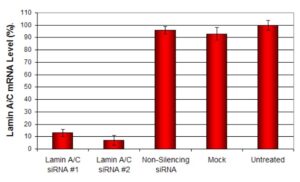
Figure: siRNAs targeting Lamin A/C mRNA or non-silencing control siRNA were transfected into fibroblast cells. At 48 hrs post-transfection the cells were analyzed by qPCR for gene expression levels and normalized to 18S rRNA levels. Values expressed as relative to the untreated sample. Data are means ± SD (n=3).
Links
Wikipedia article on fibrosis – Fibrosis (Wikipedia)
Fibroblast transfection reagent – Altogen Biosystems
Contract research laboratory RNAi Services – RNAiServices Inc
Wikipedia article on fibroblast cells – Fibroblast (Wikipedia)
Western Blot CRO Services – WesternBlotService Inc.
Tissue targeted siRNA Delivery – In Vivo Transfection Kits
CRO Pre-clinical Research Services – Xenograft animal models
Stable RNAi Cell Line Generation – RNAi Services
In Vivo siRNA Delivery – Tissue-targeted siRNA
Liposome Biology CRO – Encapsulation of Protein, RNA, and DNA Molecules into Liposomes
General Info, Cell Culture, and Transfection Information for Fibroblasts